
Computerized Tomography (CT) Scanner
|
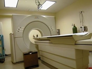
Many people have either had a CT scan or know someone who has, but you may not realize that CT is an important diagnostic tool in veterinary medicine as well. In January of 2008, we became the first veterinary clinic in Arkansas to offer in-house CT scans. Since then we have performed between 250-300 CT scans a year. In April 2013 we upgraded our CT scanner, allowing us to perform scans faster and with less anesthetic time, and some small dogs and cats can be scanned without anesthesia. Our new CT scanner also gives us the ability to build 3D reconstructions, giving us more information about your pet and the best way to help them.
The pictures below are from CT scans performed at our hospital. We hope they give you an idea of when and why CT scans are helpful to us. If you have any questions about whether CT is right for your pet, call us at 501-663-1284 and ask to speak to a doctor. |
|
Tips when viewing our CT scan pictures:
CT of the Spine: We perform CT scans of the cervical and thoracolumbar spine of dogs and cats with suspected disc extrusions, as well as less common problems such as infection of the vertebrae (diskospondylitis), vertebral and spinal cord tumors, and infection or inflammation of the spinal cord. CT scans of disc extrusions allow us to rapidly locate the disc and evaluate whether surgery is necessary. |
|
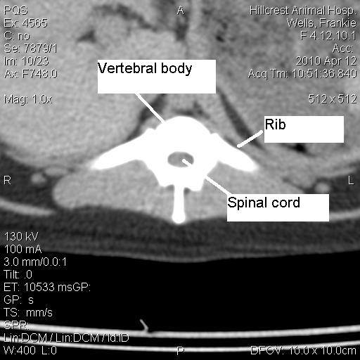
This is a normal thoracic vertebra with the spinal cord in the middle.
|
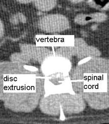
This shows a typical disc extrusion. Intervertebral disc extrusions are common in Dachshunds but may occur in any dog, at any age.
|
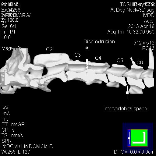
This picture is from our new CT with 3D reconstruction software showing a cervical (neck) disc extrusion.
|
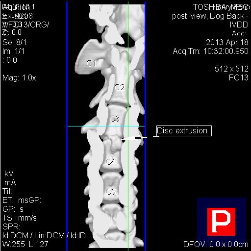
A cervical disk extrusion #2.
|
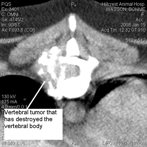
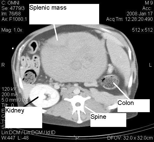
This CT scan shows a vertebral tumor eroding the vertebra. This tumor could not be seen on x-ray.
|
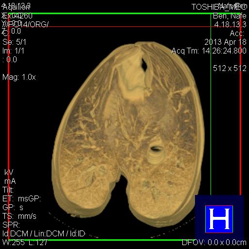
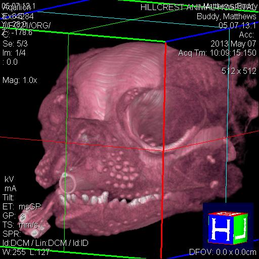
CT of the Brain: CT of the brain is often performed in dogs and cats with central nervous system (CNS) disease. We use our CT scanner to diagnose strokes, congenital diseases like hydrocephalus, trauma (skull fractures), and brain tumors. Some severe forms of infection and inflammation in the brain can also be seen on CT.
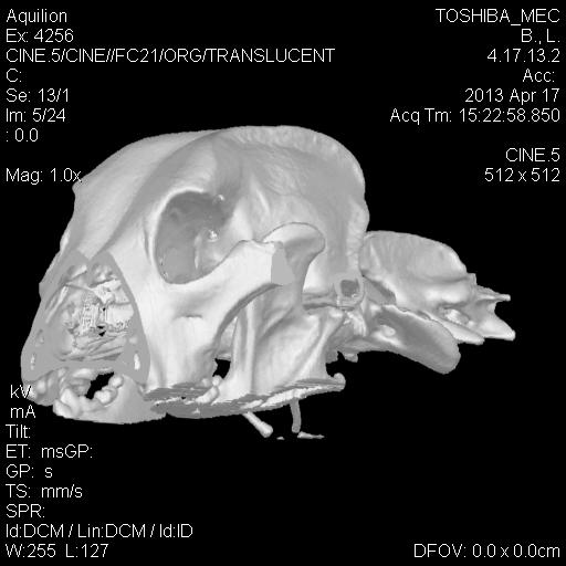
A 3D skull reconstruction.
|
A 3D skull reconstruction #2.
|
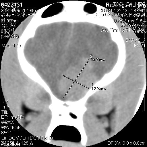
A brain tumor in 2D reconstruction.
|
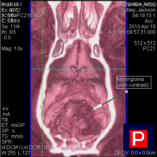
A brain tumor in 3D reconstruction.
|
|
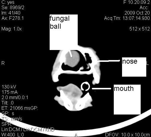
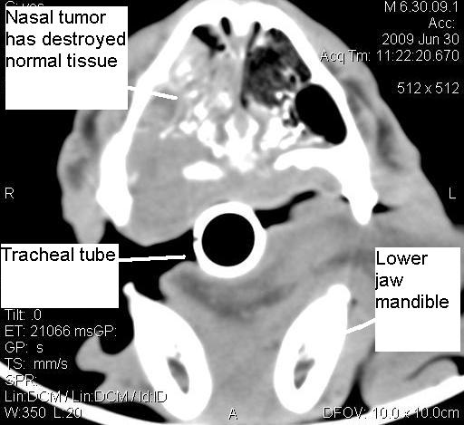
CT of the Nose: CT of the nose is indicated when dogs and cats have persistent sneezing or nosebleeds. CT scans can help us identify fungal disease, tumors, and bacterial sinusitis. Endoscopy of the nose is sometimes needed after CT to perform biopsy, a nasal flush, or a bacterial culture.
|
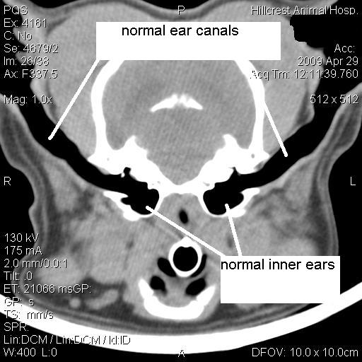
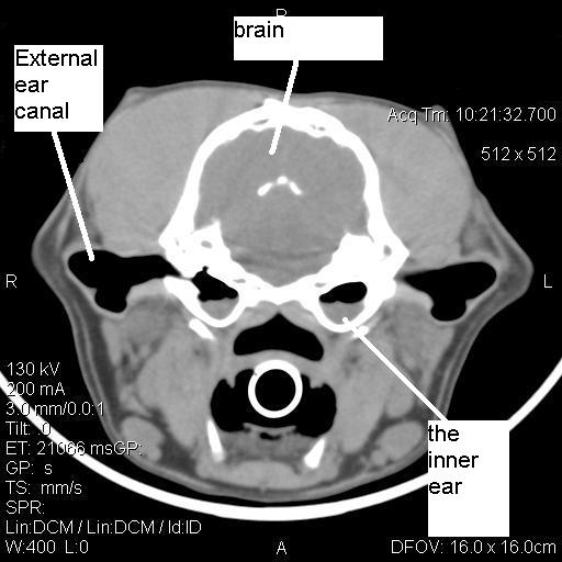
CT of the Ears: CT of the inner ears is usually performed when patients present with a head tilt or dizziness. CT can help differentiate whether the problem is due to an inner ear infection, versus a problem within the brain. In the pictures below you can see normal ear canals on the left, and, on the right, a dog with an inner ear infection in both ears.
|
|
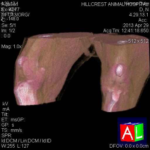
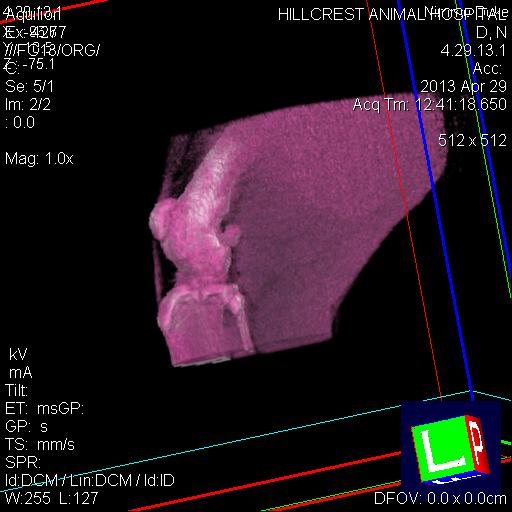
CT of the Knees and Extremities: CT scan may be performed of the knees and elbows to evaluate for bony and soft tissue changes that cannot be visualized on x-rays. Below are two 3D reconstructions of knees that were evaluated for cranial cruciate ligament rupture.
|